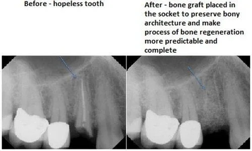
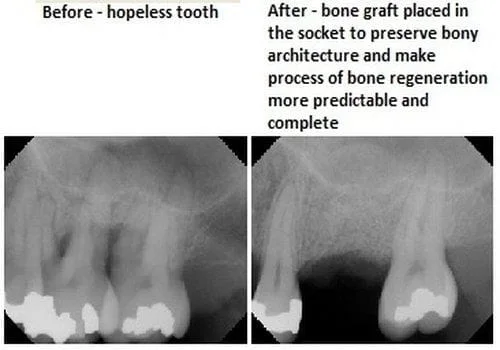
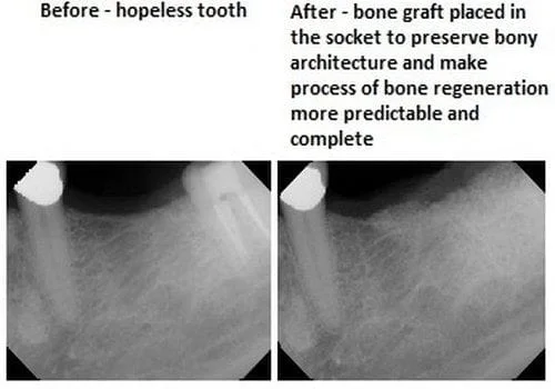
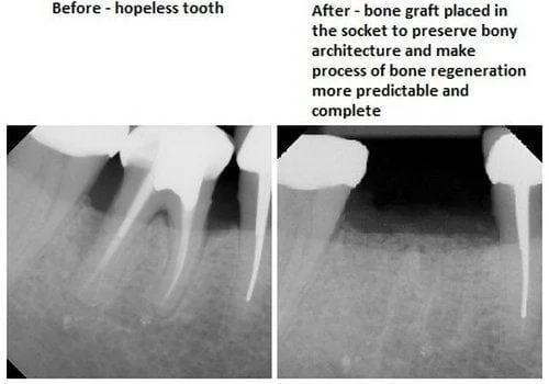
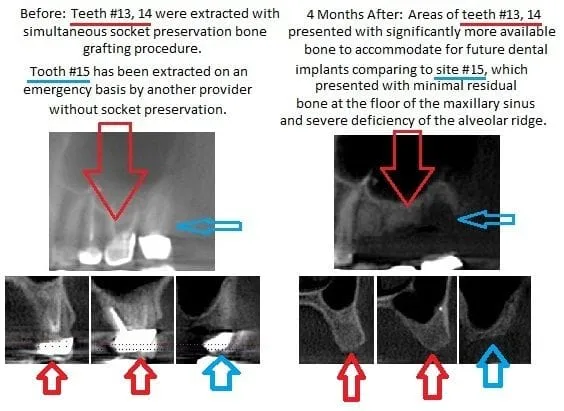
An assortment of before / after radiographs showing socket preservations completed simultaneously with tooth extractions by Dr. Pritsky.
It is important to note the change between the before and after radiographs from "dark" to "light". The "lighter" the area is, the more bone density is present. Such that, in the radiographs that go from black in the before xray to white in the after xray in a particular area, you are seeing the bone grafted into the area.
This grafted bone leads to the best possible foundation for the dental implant!
